■ 목뼈 사진은 최소한 2 가지가 필요합니다 (앞뒤면 사진, 가쪽면 사진)

이 목 척추 사진은 앞뒤면과 가쪽면 사진입니다 (AP and lateral view of cervical spine X-ray)
골절(fracture)과 탈구(dislocation)는 없습니다 (There is no fracture and dislocation found here)
또한 뼈의 정렬과 기관지앞 연부조직도 정상으로 보입니다(The alignment and pretracheal soft tissue is also looks normal)
■ 반드시 필요한 사진들 (Acquire all necessary views)
목뼈 사진 해석을 위해 필요한 사진은 3가지 입니다
(앞뒤면, 가쪽면, 치아돌기-입 벌린 면)
이제 목뼈 X-ray 해석을 위한 구조적 접근을 시작하겠습니다
(A structured approach to cervical spine X-ray interpretation is discussed below.)
■ 정확한 사진 (Adequacy)
- 관련된 모든 구조물들을 분명하게 보고 확신할 수 있도록
올바로 찍인 방사선 사진을 점검해야 합니다
(Check the radiograph's adequacy to ensure you are able to clearly all relevant structures)
■ 정렬 (Alignment)
- 3개의 선이 있습니다.
- 이 선들은 3개의 방사선 면(1 앞뒤면, 2 가쪽면, 3 치아돌기- 입 벌린 면)의 각각에서 평가하는데 필요하며,
- 이 선들은 건강한 사람들에게는 중첩되지 않습니다.
(TThere are multiple lines you need to assess across each of the three radiograph views which should run uninterrupted in healthy individuals)
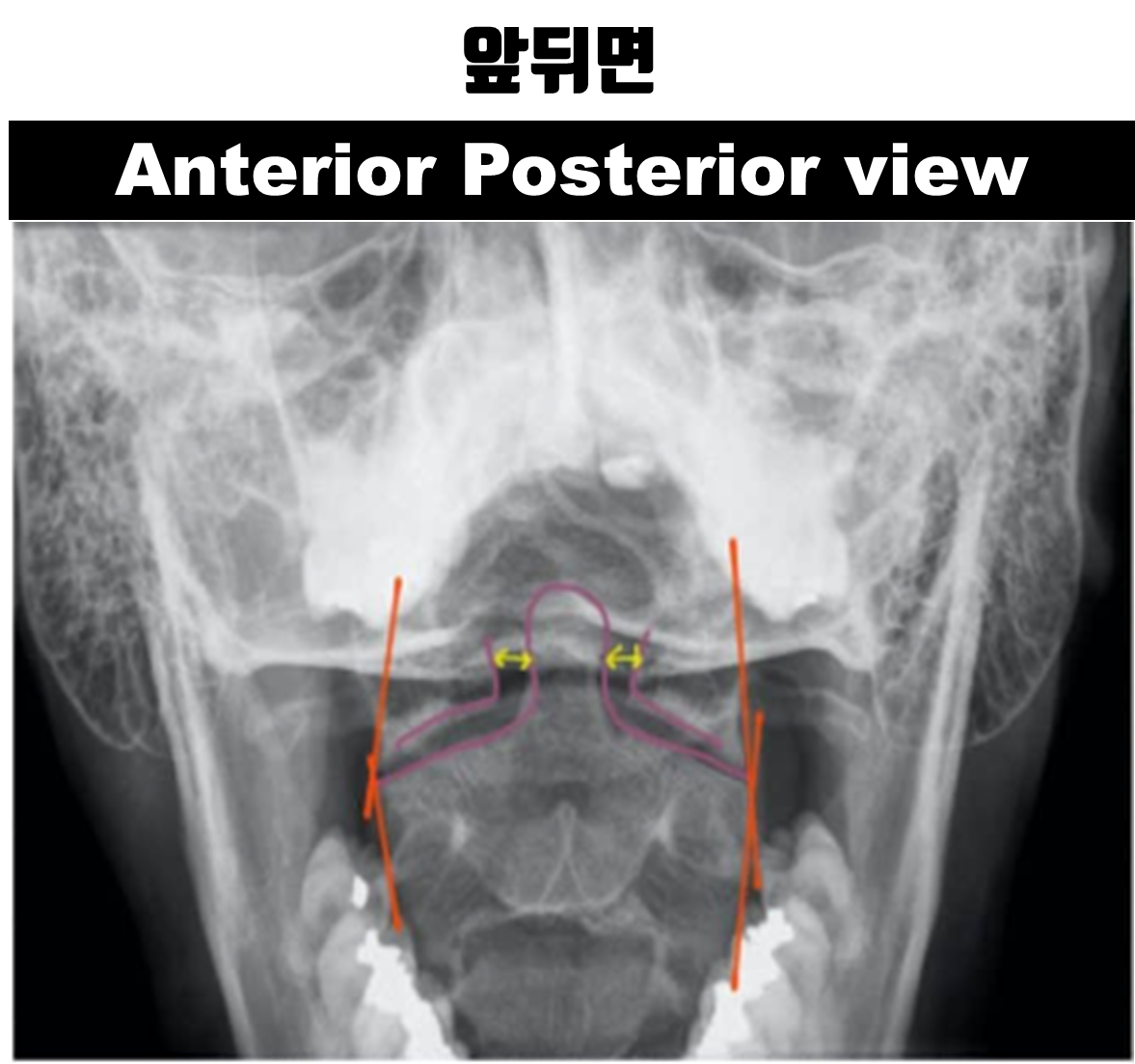
■ 1. 앞뒤면 Antero-posterior (AP) view
앞뒤면 사진에서 두 개의 가쪽 선(그림 2에서 노란색)은 척추 몸통의 양쪽을 주행합니다
(The two lateral lines of the AP view run down either side of the vertebral bodies (represented by the yellow lines in the image below).
앞뒤면 사진에서 가시돌기 선(그림 2에서 파란색)은 목뼈 1번 부터 목뼈 7번까지의 가시돌기를 잇는 선입니다
(The spinous process line runs down through each spinous process from C1 to C7 (represented by the blue line in the image below).
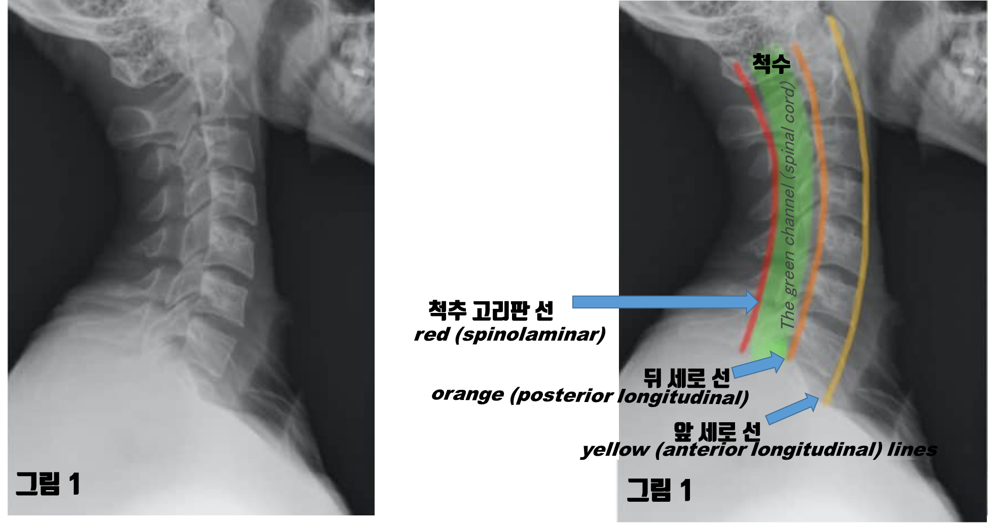
■ 2. 가쪽면 Lateral view
(The anterior longitudinal line runs along the anterior surface of the vertebral bodies)
(The posterior longitudinal line runs along the posterior surface of the vertebral bodies)
• 척추 고리판 선은 가시돌기 앞쪽 끝을 따라서 주행합니다 (가시돌기와 고리판이 관절하는 곳에서) (그림 1에서 빨간색)
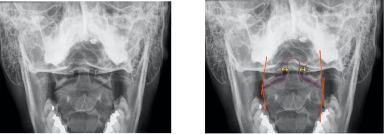
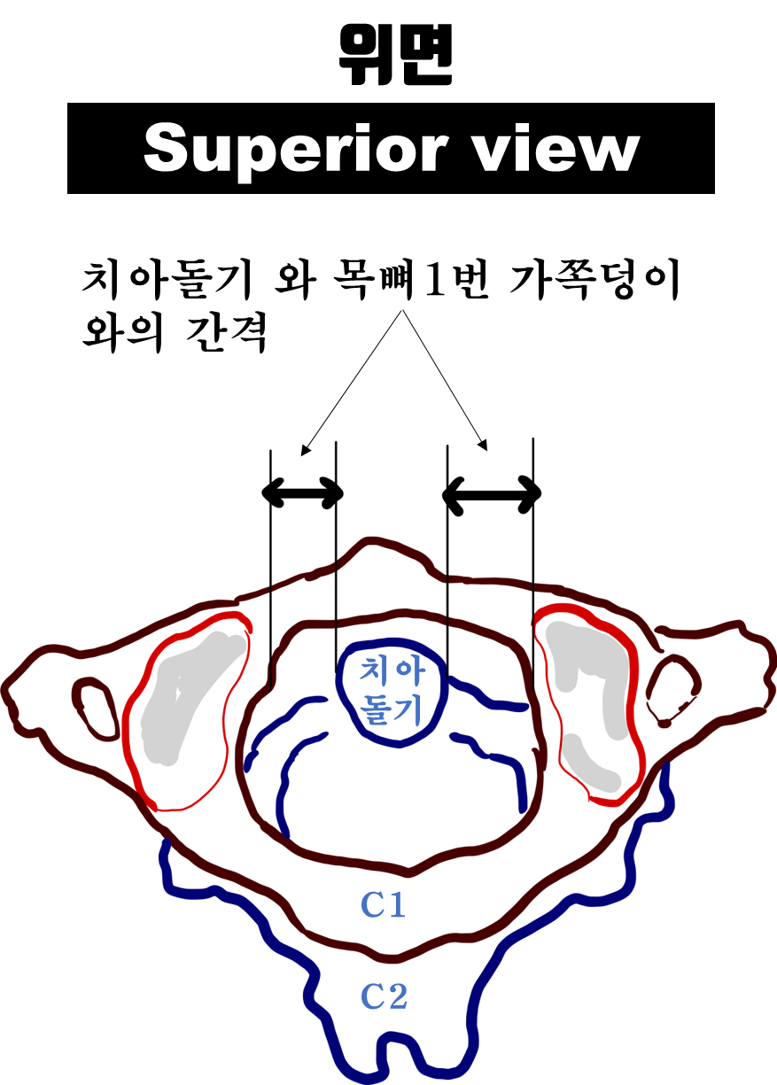
■ 3. 치아돌기- 입 벌린 면 Odontoid/open-mouth view

• 이 사진에는 몇 개의 교차하는 선이 있는데 "모통이의 만남" 이라고 언급됩니다
(The odontoid/open-mouth view has several intersecting lines which are sometimes referred to as a “meeting of corners”)
(예, 골절, 탈구)
(Irregularities in the areas where these lines intersect may indicate misalignment of the lateral masses of C1 and C2 (e.g. fracture, dislocation).)
(You can also use this view to assess the odontoid peg to make sure it is aligned with the lateral masses of C1.)
(To do this, inspect and compare the space between the peg and the lateral mass of C1 on each side)
(Asymmetry of the space between the peg and the lateral mass of C1 may indicate fracture or dislocation of the odontoid peg)
위의 사진을 위에서 본 모습은 다음과 같이 표시하면, 이 간격(위 사진에서, 노란색 부분) 을 분명하게 이해할 수 있습니다
'3_물리치료 Healing is Voltage' 카테고리의 다른 글
| https://www.aliem.com/emrad-foot-x-ray/ (0) | 2022.09.12 |
|---|---|
| 🚑 X-Ray 🚑 ; 목뼈 X-ray 해석하기 - 예제편 (0) | 2022.09.11 |
| 🚑 MRI 기초 🚑 ; T1 강조 MRI영상에서 ...지방.. 흰색으로 보여요 (0) | 2022.09.10 |
| 🚑 MRI 기초 🚑 ; 수평면, 시상면, 이마면 (0) | 2022.09.09 |
| 🚑 MRI 기초 🚑 ; 물은....T1 weighted image, T2 weighted image 기준 (0) | 2022.09.09 |


